From Guardian.co.uk:
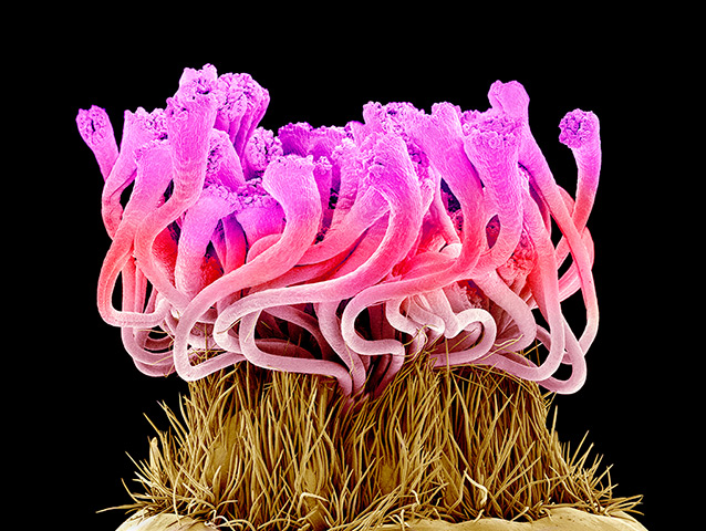
Graphic designer turned artist Susumu Nishinaga has used an electron microscope to delve deep into the fabric of petal, leaves and pollen. The Japanese artist then colours the scanning electron micrograph (SEM) images using a computer – to reveal the building blocks of life.
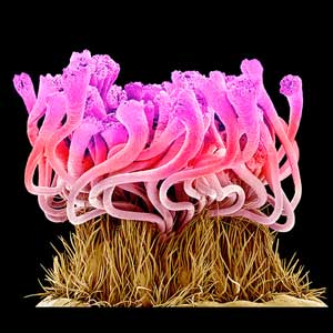
Part of the stigma (pink) of an Easter cactus flower (Rhipsalidopsis gaertneri). This is the top part of the female reproductive structure (carpel) of the flower. Pollen grains containing the male sex cells land on the stigma and may move down the style (not seen) into the ovary (not seen) Photograph: Susumu Nishinaga/Science Photo Library/ Barcroft Media


